
Incubator Accessories

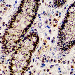
Diagnostic Biosystems BAP1 BAP1 (BAP1/2667)
BAP1 (BRCA1-Associated Protein 1) was initially identified as a protein that binds to BRCA1. BAP1 is a tumor suppressor that is believed to mediate its effects through chromatin modulation, transcriptional regulation, and possibly via the ubiquitin-proteasome system and the DNA damage response pathway. Germline mutations of BAP1 confer increased susceptibility for the development of several tumors, including uveal melanoma, epithelioid atypical Spitz tumors, cutaneous melanoma, and mesothelioma. Somatic BAP1 mutations are seen in cutaneous melanocytic tumors (epithelioid atypical Spitztumors and melanoma), uveal melanoma, mesothelioma, clear cell renal cell carcinoma, and other tumors. BAP1 immunohistochemistry is particularly useful in differentiating malignant mesothelioma (nuclear negative) vs. reactive mesothelial proliferation (nuclear positive).

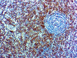
Diagnostic Biosystems CD7 CD7 (CBC.37.80)
CD7 is expressed by most peripheral blood T cells, NK cells, and all thymocytes. It is one of the earliest surface antigens on T and NK-cell lineages. The antibody is a useful aid for classification of T-cell malignancies.
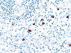
Diagnostic Biosystems IGM IgM
This antibody reacts with the heavy chain present in all types of human IgM. It stains B cell follicles in human lymphoid tissues. In lymphoid tissues, IgM containing plasma cells stain strongly but immune complexes are usually undetectable.
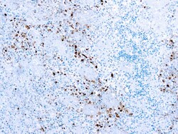
Diagnostic Biosystems HSV I Herpes Simplex Virus Type I (HSV1)
This antibody reacts with HSV type I specific antigens and with antigens common to HSV type I and II virus. It reacts with all the major glycoproteins present in the viral envelope as well as with at least one core protein.
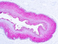
Diagnostic Biosystems CYTOKERATIN WIDE SPECTRUM Cytokeratin Wide Spectrum
This antibody reacts with cytokeratins of 58, 56, 52, 60, 51, 48 and 68 kDa MW. This antibody is well suited for the staining of a broad spectrum of human keratins.
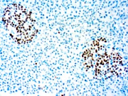
Diagnostic Biosystems INSM1 A-8 INSM1 (A-8)
INSM1 (insulinoma-associated protein 1), also known as zinc-finger protein IA-1, is a developmentally regulated zinc-finger transcription factor. It localizes to the nucleus and is expressed in embryonic tissues undergoing neuroendocrine differentiation. INSM1 is not expressed in normal adult tissues but it can be found highly expressed in neuroendocrine tumors. INSM1 contains five Cys2-His2-type zinc-finger DNA binding domains and a prohormone domain. INSM1 acts as a transcriptional repressor of the Neuro D promoter and recruits cyclin D1 as a corepressor. It plays an importantrole in neuroendocrine development and isrequired for normal differentiation of pancreatic endocrine cells. Inhibition of INSM1 results in decreased formation of glucagon and Insulin positive cells. The gene encoding INSM1 is directly regulated by Neurogenin 3 which binds chromatin in the INSM1 promoter region and induces transcription.
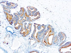
Diagnostic Biosystems P504S p504S/α-Methylacyl-CoA Racemase (AMACR)
P504S has been recently described as a prostate cancer-specific gene that encodes a protein involved in the β-oxidation of branched chain fatty acids. Expression of p504S protein is found in prostatic adenocarcinoma but not in benign prostatic tissue. Itstains premalignant lesions of prostate: high-grade prostatic intraepithelial neoplasia (PIN) and atypical adenomatous hyperplasia. P504S can be used as a positive marker for PIN.
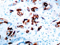
Diagnostic Biosystems MAMMAGLOBIN A Mammaglobin A
The mammaglobin gene was first identified using a differential screening approach directed at the isolation of novel, human breast cancer-associated genes. Mammaglobin-A is highly overexpressed in breast cancer cell lines and primary breast tumors. This pattern of expression is restricted to mammary epithelium and metastatic breast tumors. Thus, mammaglobin-Aspecific T cell immune responses may provide an important approach for the design of breast cancer-specific immunotherapy.
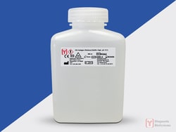
Diagnostic Biosystems 10X ANTIGEN RETRIEVAL 10X Antigen Retrieval Buffer High pH 10.0
10X Antigen Retrieval Buffer High pH 10.0 is designed for use during the heat induced epitope retrieval (HIER) step prior to immunohistochemistry on formalin-fixed paraffin embedded tissue sections. The use of this buffer in combination with heat (often by microwave, water bath, or pressure cooker) has been shown to restore the antigenicity of proteins modified during the formalin fixation of tissue. This buffer is supplied as a 10X stock solution.
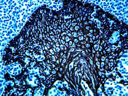
Diagnostic Biosystems PERMABLACK/HRP PermaBlack/HRP
PermaBlack/HRP is a substrate-chromogen system designed to be used for either IHC or ISH when utilizing horseradish peroxidase. PermaBlack/HRP can be permanently mounted to produce a sharply contrasting ebony black color that can be easily distinguished from other stains.

Diagnostic Biosystems ALK PHOS ENHANCER Alkaline Phosphatase Enhancer
This product is used as a signal enhancer in conjunction with alkaline phosphatase based immunostaining systems.\n\nAlkaline Phosphatase, an enzyme derived from bovine intestinal mucosa, is often used as a label for in situ hybridization, immunohistochemistry, Southern and Northern hybridization and DNA sequencing. Detection of this enzyme requires the generation of an insoluble colored reaction end product. Stronger the signal, better the staining. DBS introduces a new stable solution to increase the signal generated by alkaline phosphatase several fold. A single treatment of tissue sections with enhancer before the treatment with substrate/chromogen enables the visualization of difficult to localize antigens. This enhancer works well with Fast Red but can be used with BCIP/NBT and BCIP/INT also. Specimens stained with the above substrates/chromogens can not be dehydrated in ethanol and hence should be mounted in aqueous based mounting medium like C/C Mount (DBS catalog # K002).
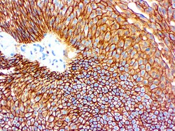
Diagnostic Biosystems CD44 H CAM CD44 /HCAM
This antibody is specific to a cell surface glycoprotein of 80‐95 kDa, known as CD44. CD44 is expressed on the surface of lymphocytes, monocytes and granulocytes. CD44 immunostaining is commonly used for the discrimination of urothelial transitional cell carcinoma in-situ from non-neoplastic changes in the urothelium.
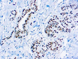
Diagnostic Biosystems WILMS TUMOR 1 PROTEIN WT1 Wilm’s Tumor 1, WT1
Wilm’s Tumor (WT1), is a gene involved in the induction of Wilm’s Tumor, a sporadic and familial pediatric kidney tumor. Wilm’stumor is associated with mutations of WT1, a zinc-finger transcription factor that is essential for the development of the metanephric kidney and the urogenitalsystem. The WT1 gene is normally expressed in fetal kidney and mesothelium, and its expression has been suggested as a marker for Wilm’s tumor and mesothelioma. WT1 has been identified in proliferative mesothelial cells, malignant mesothelioma, ovarian carcinoma, gonadoblastoma, nephroblastoma, and desmoplastic small round cell tumor.
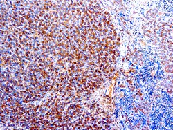
Diagnostic Biosystems ALPHA-1-ANTITRYPSIN Alpha (α )-1-Antitrypsin
The antibody isspecific against human α-1-antitrypsin. This antibody stains macrophages in human tissues and does not stain any other cell type.
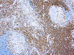
Diagnostic Biosystems CD3 CD3, T Cell
This antibody reacts with intracytoplasmic portion of the CD3 antigen on the T cells. It stains T cells in cortex as well as medulla of the thymus and lymphoid tissues. This antibody also labels T cell neoplasm, malignant histiocytes and Reed-Sternberg cells in Hodgkin disease.